Thursday 28 May 2020

LUNGS

LUNGS : 

 
  • How to Breathe - Thrive Physical Therapy
  • The lungs occupying major portion of the thoracic cavity , leave little space for the heart ,which excavates more of the left lung .  
  • The two lungs hold the heart tight between them, providing it the protection it rightly deserves. 
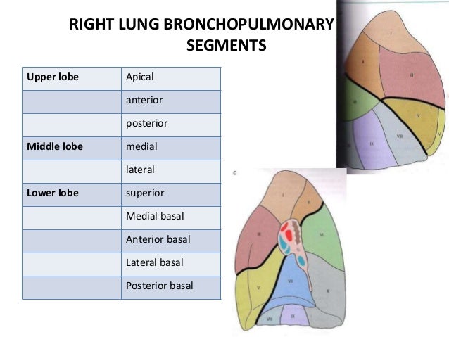
  • There are each bronchopulmonary segments in each lung. 
  • The lung are a pair of respiratory organs situated in the thoracic cavity. 
  • Each lung invaginates the corresponding pleural cavity. 
  • The right and left lung is separated by the mediastinum.  
  • Lungs are spongy in the texture . 
  • In the young's the lungs are brown or grey in colour. 
  • Gradually , they become mottled black because of the deposition of  inhaled carbon particles. 
  • Medical Education Chart of Biology for Lungs Diagram. Vector ...

Measurements : 
  • The right lung weight about 700 g .
  • It is about 50 to 100 g heaver than the left lung. 
Features :  Each lung is conical in shape . it has : 
  • An apex at upper end .
  • A base resting on the diaphragm.
  • Three borders, i.e. anterior, posterior , and inferior. 
  • Two surfaces i.e. costal and medial. The medial surface is divided into vertebral and mediastinal parts. 
APEX :  The apex is blunt and lies above the level of the anterior end of the first rib. It reaches nearly 2.5 cm above the medial one third of the clavicle, just medial to the  supraclavicular fossa. It is covered by the cervical pleura and by the suprapleural membrane, and is grooved by the subclavian artery on the medial side and anteriorly.

BASE : The base is semilunar and concave. It rests on the diaphragm which separates the right lung from the right lobe of the liver , and left lung from the left lobe of the liver, The fundus of stomach  and spleen. 
 
BORDERS : 
  1. Anterior border : The anterior border is very thin . It is short than the posterior border. On the right side, it is vertical and corresponds to the anterior or costomediastinal line of pleural reflection. The anterior border of the left lung shows a wide cardiacnotch below the level of the fourth costal cartilage. 
  2. Posterior : The posterior border is thick and ill defined. it corresponds to the medial margins of the heads of the ribs. it extends from the level of the seventh cervical spine to the tenth thoracic spine. 
  3. Inferior Border : The inferio border separates the base form the costal and medial surfaces. 
SURFACES : 
  1. Costal surface : The costal surface is large and convex . It is in contact with the costal pleura and the overlying thoracic wall . 
  2. Medial surface : The medial surface is divided into a posterior or vertebral part, and an anterior or mediastinal part.  
Fissures and lobes of the lungs : 
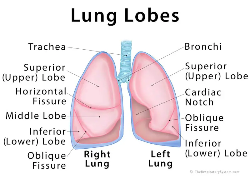
  •    The right lung is divided into 3 lobes ( upper , middle, lower ). By two fissures , Oblique and horizontal. The left lung is divided into two lobes by the oblique fissure. 
Lung Lobes: Definition, Anatomy, Functions, Picture

RELATIONS OF LUNGS : Lung anatomy.


DIFFERENCE BETWEEN RIGHT AND LEFT LUNG : 


Difference Between Right and Left Lung | Compare the Difference ...
Difference Between Right and Left Lung


Clinical significance


Wednesday 27 May 2020

HEART

HEART : 
 Computer generated animation of a beating human heartHeart anterior exterior view.jpg
Features : 
  • Heart is a conical hollow muscular organ situated in the middle mediastnum. 
  • It is enclosed within the pericardium .
  • It pumps blood to various parts of the body to meet their nutritive requirements .
  • The greek name for the heart is cardia from which we have the adjective cardia. 
  • The latin name for the heart is cor from which we have the adjective coronary. 
  • The heart is placed obliquely behind the body of the sternum and adjoining part of the costal cartilages, so that one-third of it lies to the right and two-third to the left median plane. 
  • Measurements : The heat measures about 12 ✖ 9 cm and weight about 300 g in males and in females 250 g in female 

External Features : 
  • The human heart has four chambers . 
  • These are the right and left atria and right and left ventricles. 
  • The atria lie above and behind the ventricles. On the surface of the heart , they are separated from the by ventricle an atrioventricular groove. 
  • The atria are separated from each other by an  interatrial groove. 
  • The ventricles are separated from each other by an interventricular groove, which is subdivided into anterior and posterior parts . 
The heart has : 
  • An apex directed downwards, forwards and to the left,
  • A base (posterior surface) directed backwards 
  • Anterior / sternocoastal
  • Inferior and lateral surfaces. 
The surfaces are demarcated by upper, inferior, right and left borders . 

Chambers : 
Valves : 
  • The heart has four valves, which separate its chambers. One valve lies between each atrium and ventricle, and one valve rests at the exit of each ventricle.
  • The valves between the atria and ventricles are called the atrioventricular valves.  
  • Between the right atrium and the right ventricle is the tricuspid valve
  • The tricuspid valve has three cusps,which connect to chordae tendinae and three papillary muscles named the anterior, posterior, and septal muscles, after their relative positions. The mitral valve lies between the left atrium and left ventricle.
  •  It is also known as the bicuspid valve due to its having two cusps, an anterior and a posterior cusp.

Heart wall : 


Pericardium

  • The pericardium is the sack that surrounds the heart. 
  • The tough outer surface of the pericardium is called the fibrous membrane. 
  • This is lined by a double inner membrane called the serous membrane that produces pericardial fluid to lubricate the surface of the heart.


Tuesday 26 May 2020

Bronchopulmonary Segment

Bronchopulmonary Segment :

Intro : 
  • Each Tertiary bronchus enters into the one bronchopulmonary segment of the lungs .
  • These are well defined anatomic, functional, and surgical sector of the lung.
  • A bronchopulmonary segment is independent unit of the lung. 
  • i.e. is bronchopulmonary segment is aerated by an indepent segmental bronchus, supplied by independent branch of pulmonary artery but the pulmonary vein is intersegmental .
  • Lung Anatomy Bronchopulmonary Segment
  • bronchopulmonary segment is a portion of lung supplied by a specific segmental bronchus and arteries.
  • These arteries branch from the pulmonary and bronchial arteries, and run together through the center of the segment.
  • The segments are separated from each other by layers of connective tissue that forms them into discrete anatomical and functional units.
  • There are ten bronchopulmonary segment in each lung .
  • It is very important to know about the bronchoulmonary segment whenever they are diseased , only the affected bronchopulmonary segment can be removed . 
  • Each bronchopulmonary segment is divided into many lobules by connective tissue . 
  • bronchopulmonary segments - Google Search | Bronchopulmonary ...
Right Bronchopulmonary Segment : There are ten bronchopulmonary segments in the right lung : 
 
  • Right lung

    • Superior lobe.
      • apical segment.
      • posterior segment.
      • anterior segment.
      • Bronchopulmonary segments
    • Middle lobe. 
      • lateral segment.
      • medial segment.
    • Inferior lobe.
      • superior segment.
      • medial-basal segment.
      • anterior-basal segment.
      • lateral-basal segment.  
      • posterior-basal segment.

Left Bronchopulmonary Segment : Some of the segments may fuse in the left lung to form usually eight to nine segments (four to five in the upper lobe and four to five in the lower lobe.

  • Left lung : 

    • Superior lobe
      • apico-posterior segment (merger of "apical" and "posterior")
      • anterior segment
    • Lingula of superior lobe
      • inferior lingular segment
      • superior lingular segment 
      • Bronchopulmonary segments
    • Inferior lobe
      • superior segment
      • anteromedial basal segment (merger of "anterior basal" and "medial basal")
      • posterior basal segment
      • lateral basal segment


Clinical Anatomy : 
  • Usually the infection of the bronchopulmonary segment remains restricted to it, although tuberculosis and bronchogenic carcinoma may spread from one segment to another.
  • Visualising the interior of the bronchi through a bronchoscope passed through the mouth and trachea, procedure is called bronchoscopy.
  • The carina of the trachea is a hook shaped process projecting backward from the lower margin of lowest tracheal ring. It helps to divide trachea into two primary bronchi. Right bronchus makes an angle of 25°, while left one makes an angle of 45°.
  • Apical segment of lower lobe is the most dependent bronchopulmonary segment in supine position .Foreigen bodies are likely to be lodged here . 


Sunday 24 May 2020

Blood Circulation of Kidney ( Renal Circulation )

Blood Circulation of Kidney ( Renal Circulation )

  • The renal circulation supplies the blood to the kidneys via the renal arteries, left and right, which branch directly from the abdominal aorta. Despite their relatively small size, the kidneys receive approximately 20% of the cardiac output
  • Blood vessels of kidneys are highly specialised to facilitate the function of nephrons in tjhe formation of urine. 


  • Each renal artery branches into segmental arteries, dividing further into interlobar arteries, which penetrate the renal capsule and extend through the renal columns between the renal pyramids. The interlobar arteries then supply blood to the arcuate arteries that run through the boundary of the cortex and the medulla. Each arcuate artery supplies several interlobular arteries that feed into the afferent arterioles that supply the glomeruli
What is the correct flow of blood in the kidneys? - Quora
  • After filtration occurs, the blood moves through a small network of venules that converge into interlobular veins. As with the arteriole distribution, the veins follow the same pattern: the interlobular provide blood to the arcuate veins then back to the interlobar veins, which come to form the renal vein exiting the kidney for transfusion for blood.


Saturday 23 May 2020

Eye (Visual Pathway)


Eye : 

Eyes are organs of the visual system. They provide animals with vision, the ability to receive and process visual detail, as well as enabling several photo response functions that are independent of vision. Eyes detect light and convert it into electro-chemical impulses in neurons

Visual Pathway / Optic pathway : 
  •     Visual pathway or optic pathway is the nervous pathway that transmit the impulses from the retina visual center to the cerebral cortex. 
  • In binocular vision , the light rays from the temporal half of visual field fall upon the nasal part of the corresponding retina . 
  • The ray from the nasal half of visual field fall upon the temporal part of the retina . 
COURSE OF VISUAL PATHWAY : Visual pathway consists of the six components : 
  1. Optic nerve.
  2. Optic chismata .
  3. Optic tract.
  4. lateral eniculate body . 
  5. optic radiations.
  6. Visual cortex. 

Human visual pathway.svg
1.Optic nerve : 
  • optic nerve is formed by the axon of the ganglionic cell optic nerve leaves the through the optic disk.
  • Both the fibers of nasal half part and temporal half part of retina carry the impulses from temporal half and nasal half of visual field of the same eye.
2. Optic chiasma. : 
  • Medial fiber of each optic nerve cross the mid line and join the uncrossed lateral fibers of the opposite side . to from the Optic tract . This area of the optic nerve fibers is called optic chiasma. 
3.  Optic tract. : 
  • Optic tract is formed by uncrossed fibers of optic nerve on the same side and crossed fibers of optic nerve from opposite side . 
  • All fibers of optic tract runs backwards, outwards, towards the cerebral peduncle. 
4 . Lateral geniculate body . 
  • Majority of thefibers of optic tract terminate in lateral geniculate body . Which form subcortical centere for visual sensation . 
  • From here geniculocalcarine tract or optic radiation arises . this tract is the last relay of visual pathway. 
  • Visual pathway in a primate. The superior colliculus, lateral ...
5. Optical radiations : 
  • Fibers from the lateral geniculate body pass through internal capsual and from optic radiation .
  • The fibers between lateral geniculate body and visual cortex are also called geniculocalcarine fibers. 
  • Optic radiations end in the visual cortex. 
6. Visual cortex :
  • Primary corticalcenter for vision is called visual cortex. Which is located on the middle surface of occipital lobe .
Area of isual cortex and their functions : Three area resent in the visual cortex ,
  1. Primary visual area (area 17) which is concerned with the percepton of visual impulses . 
  2. Secondary visual area or visual association area (area 18 ) which is concerned with the interpretation  of visual impulses . 
  3. Occipital eye field (area 190 which is concerned with the movment of eyes. 


Friday 22 May 2020

NEPHRON

                                                       NEPHRON   

Introduction : 
Nephron anatomy with labeled
  • Nephron is defined as functional unit of the kidney . 
  • Each kidney consists of 1 to 1.3 millions of nephrons. 
  • The number of nephrons starts decreasing after about 45 to 50 years of age at the rate of 0.8 to 1 % every year. 
  • Each nephrons is formed by two parts :
  1. A blind end called renal corpuscle or malpighian corpuscle .
  2. A tubular portion called renal tubule.
RENAL CORPUSCLE : 
  • Renal corpuscle or Malpighian corpuscle is a speroidal and slightly flattened structure with a diameter of about 200 Âµ.
  • Function of the renal corpuscle is the filtration of lood which form the first phase of urine formation.  
  •  
STRUCTURE OF RENAL CORPUSCLE AND MALPIGHIAN CORPUSCLES : 
  Renal corpuscle is formed by the two portions : 
  1. Glomerulus .
  2. Bowman capsule.   What is the Bowman's capsule? What are the main functions? - Quora
 1. Glomerulus : Glomerulus  is a tuft of capillaries enclosed by Bowman capsule. It consists of glomerular capillaries interposed afferent arteriole on one end and efferent arteriole on the other end . 

Function : 
   Glomerular capillaries are made up of single layer of endothelial cells, which attach to the basement membrane . Endothelium has many pores called the filtration pores . this presence shows the filtration is the functuion of the glomerulus.

2. Bowman Capsule :  Bowman capsule is a capsular structure, which encloses the glomerulus . 
         It is formed by the two layer : (a) Inner visceral layer , (b) Outer parietal layer. 
   Visceral layer covers  the glomerular capillaries . It is continued as the parietal layer at the visceral pole . 
 
Function : 
        Functional anatomy of the bowman capsule resembles a funnel with filter paper. Diameter of bowmans capsule is 200µ .

 TUBULAR PORTION OF THE NEPHRON :
 
  Tubular [portion of the nephron is continuation of the Bowman capsule .
         It is made up of three parts : 
  1. Proximal convulated tubule  (PCT ). 
  2. Loop of the Henle.
  3. Distal convulated tubule (DCT) . 

  1. Proximal convulated tubule  (PCT ).  : Proximal convulated tubule is the coiled porion arising from the Bowman capsule . It is situated in the cortex . It is continued as descending limb of loop of Henle.  length of proximal convulated tubule is 14 mm and diameter is 55 Âµ .   Proximal convulated tubule is continued  as Loop of Henle. 
  2.  Loop of Henle :  Loop of Henle is consists  of :  (a) Descending limb (b) Hairpin Bend   (c) Ascending limb. 
  •  (a) Descending limb : Descending limb of   Loop of Henle is made up of two segments,  i.e. is (1) Thick descending segment (2) Thin descending segment .  
           (1) Thick descending segment : Thick descending segment is direct continuation of the Proximal convulated tubule. It is descent into medulla.  
            (2) Thin descending segment : Thick descending segment is continued as thin descending segment . And it is continued as the hairpin bend of the loop .

The Excretory System: Functional Anatomy (MCAT 2018: Biology ...
  • (b) Hairpin Bend :   Hairpin Bend formed by the flattened epithelial cells without brush border and it is continued as the ascending limb of loop of the Henle. 
  •      Ascending limb : Ascending limb made up of two parts , (1) Thick ascending segment (2) Thin ascending segment . The Thick ascending segment ascends to the cortex and continues as distal convulated tubule . 
 3. Distal convulated tubule (DCT) Distal convulated tubule is continuation of the thick ascending segment and occupies the cortex of the kidney. It continues s a collecting duct . The length distal convulated tubule is 14.5 to 15 mm It has diameter of 22 to 50 Âµ. 
 
Collecting duct : 
Distal convulated tubule is continues as the intial or arched collecting duct , which is in cortex. The lower part of the collecting duct is lies in the medulla. Length of the collecting duct is 20 to 22 mm and its diameter is 40 to 200 Âµ . 


Wednesday 20 May 2020

Ear ( Structure of Ear )

Structure of Ear : 
The ear is the organ of hearing and, in mammals, balance. In mammals, the ear is usually described as having three parts—the outer ear, the middle ear and the inner ear

Human Ear: Structure and Anatomy - Online Biology Notes

Outer Ear : 
  • The outer ear is the external portion of the ear and includes the fleshy visible pinna (also called the auricle), the ear canal, and the outer layer of the eardrum (also called the tympanic membrane). 
  • Pinna : The flap like structure on either side of head is the Pinna. It is made up of elastic fibrocartilage covered by skin that is why it is very flexible. Soft lower projecting part is the lobule of the ear . 
  • External auditory meatus. (ear canal) : From the external opening on the lateral side of the head to the tympanic memebrane (ear drum ) extents as a "S" shaped tube known as the external meatus. It is about 26 mm long and 7 mm in diameter . It is lateral 1/3rdb  is cartilaginous and medial 2/3rd is bony. The external auditory meatus is internally lined by the skin having numerous ceruminous glands. ( wax producing glands )   and hair. 
  • Congenital Ear Deformity Microtia and Anotia - EarWell Centers of ...
  • Tympanic membrane : Tympanic membrane seprates the external ear from the middle ear. The surface of tympanic membrane towards the external auditory meatus is lined by the skin . The surface of the tympanic membrane towards the middle ear is lined by the mucous membrane . In between the two layer is a fibrous tissue.  Malleus and incus are releted here on the medial side of tympanic membrane.

Middle Ear ( Tympanic cavity )  : 
  • The middle ear lies between the outer ear and the inner ear.
  •  It consists of an air-filled cavity called the tympanic cavity and includes the three ossicles and their attaching ligaments; the auditory tube; and the round and oval windows.
  • The middle ear also connects to the upper throat at the nasopharynx via the pharyngeal opening of the Eustachian tube.
  • It is dumbbell shaped air filled cavity in the petrous part of temporal bone . 
  • It has roof, a floor, an anterior wall , a posterior wall ,a medial wall , and lateral wall.
  • Roof : Pectorus part of temporal bone 
  • Floor : Petrous part of temporal bone.
  • Anterior wall : Petrous part of temporal bone in which opens the  auditory tube . this tube maintaining same air pressure on both the sides of tympanic membrane .
  • Posterior Wall : Air filled cavity in the pectrous part of temporal bone known as the mastoid antrum . 
  • Lateral wall : Tympanic membrane is present on that side. 
  • Midde ear is linned by the mucous membrane and three small bones i.e. malleus , incus , stapes .
  • The ossicles are three small bones that function together to receive, amplify, and transmit the sound from the eardrum to the inner ear.
  • The ossicles are the malleus (hammer), incus (anvil), and the stapes (stirrup). 
  • The stapes is the smallest named bone in the body.
  • The three ossicles transmit sound from the outer ear to the inner ear. The malleus receives vibrations from sound pressure on the eardrum, where it is connected at its longest part (the manubrium or handle) by a ligament. It transmits vibrations to the incus, which in turn transmits the vibrations to the small stapes bone. The wide base of the stapes rests on the oval window. As the stapes vibrates, vibrations are transmitted through the oval window, causing movement of fluid within the cochlea.
Inner Ear : 
  • The inner ear sits within the temporal bone in a complex cavity called the bony labyrinth.
  • A central area known as the vestibule contains two small fluid-filled recesses, the utricle and saccule.
  • There  is a bony labyrinth inside which is membranous labyrinth . It lies in the pectoral part of the temporal bone . 
  • The membranous labyrinth is filled with the fluid called endolymph. 
  • Bony labyrinth consist of three parts, 
  1. Cochlea ( Anteriorly ) 
  2. Vestibule (Middle )
  3. Semicircular canal ( Posteriorly ) . 
  • Membranous labyrinth : It lies within bony labyrinth separated by watery fluid known as perilymph. inside the membranous labyrinth is watery fluid known as endolymph . Vibrations are transmitted into the inner ear into a fluid called endolymph, which fills the membranous labyrinth. 
Functions : 
  1. Hearing : Sound waves travel through the outer ear, are modulated by the middle ear, and are transmitted to the vestibulocochlear nerve in the inner ear. This nerve transmits information to the temporal lobe of the brain, where it is registered as sound. 

     
  2. Balance : Providing balance, when moving or stationary, is also a central function of the ear. The ear facilitates two types of balance: static balance, which allows a person to feel the effects of gravity, and dynamic balance, which allows a person to sense acceleration.


Tuesday 19 May 2020

Relations of Kidneys

Relations of Kidneys  :  

We will be learn in the previous blog about the all basic information about kidney , if you want read it for references click on the  kidney is in blue colour KIDNEY . 
Kidney
Relations of Kidneys 
  • The kidneys are the retroperitonial organs and are only partly covered by peritoneum anteriorly. There are two kidneys which divide into three types of Superiorly , Anteriorly, & Posteriorly .  Relations are as below, 
 Relations  Right KidneyLeft kidney 
 Superiorly   Right adrenal gland Left adrenal gland  
 AnteriorlyRight suprarenal gland ,
Right lobe of the liver,
Duodenum ,
Hepatic fiexure of colon ,
Small intestine. 
 Left suprarenal gland, 
Spleen, 
Stomatch,
Pancrease, 
Jejunum .
 Posteriorly

Diaphram,
Muscle of posterior abdominal wall,
12th Ribs,
Subcostal vesseles ,
Iliohypogastric nerve. 
 Diaphram,
Muscle of posterior abdominal wall,
11th,12th Ribs,
Subcostal vesseles ,
Iliohypogastric nerve. 
  

JaypeeDigital | eBook Reader

Monday 18 May 2020

Nerve Supply Of Tongue

Nerve Supply Of Tongue . 

  The tongue is a muscular organ situated in the floor of the mouth . It associated with the function of  Taste,  Speech, chewing, deglutition . Tongue comprises skeletal muscle which is voluntary. 
There are two types of nerve supply to the tongue. Which are as follow,

The Tongue & it's Innervation!

Motar Nerves : 
  •  All intrensic and extrensic muscles, except palatoglossus, are supplied by the hypoglossal nerve.
  • The palatoglossus is supplied by the cranial root of the accessory nerve through the pharyngeal plexus .  
  • So seven out of eight muscles are supplied by Xll nerve. 

Sensory nerve : 
  •  The lingual nerve is nerve of general sensation, and chorda tympani is the nerve for the anterior 2/3 of the tongue except vallate papillae.
  • The glossopharyngeal nerve is the nerve for both general sensation and taste for the posterior 1/3 of the tongue including the circumvallate papillae. 
  • The posterior most of the tongue is supplied by the vagus nerve through the internal laryngal branch.  
  • anatomy of oral cavity ,tongue and palate

Sunday 17 May 2020

Papillae of Tongue

Papillae of Tongue  : 

     The tongue is a muscular organ situated in the floor of the mouth . It associated with the function of  Taste,  Speech, chewing, deglutition . Tongue comprises skeletal muscle which is voluntary.
   Papillae are projections of mucus membrane or corium which gives the anterior (2/3) two - thrid of the tongue. Its has characteristic roughness . 
List of Papillae of Tongue – Location and Histology

These are following three types : 
  1. Vallate or circumvalate papillae : They are large in the size about 1-2 mm in diameter and 8-12 in number. They are situated in the front of sulcus terminalis. Each papillae is cylindrical projection surrounded by a circular sulcus. The wall of papillae have taste buds.  Anatomy of Taste, Illustration
  2. The fungiform papillae : They are numerous near the tip and margin of the tongue . But some of them scattered over the dorsum . These are smaller than vallate papillae but larger than filiform papillae. They are distinguished by bright red colour. 
  3. The filiform papillae or conical papillae : They are cover the presulcal area of the dorsum of tongue and give it a valvety apperance . They are smallest and most numerous of the lingual papillae.
  4. Few palliate papillae are aslso present . 

Glands of Skin

Glands of Skin

    Skin is largest organ of body. It is outer most covering of human body . It protect the underling muscles, bone, ligaments and internal organs.It is composed of the superficial layer the epidermis and a layer deeper to it known as dermis . 
    Two types of glands are present in the skin over most of the body . These are sweat gland and sebaceous glands. 

What Are Sweat Glands? - Understanding Infected Sweat Glands ...
Skin glands

Sweat Gland : The sweat gland and their duct derived epidermis . They are abundance in palm, sole axilla, groins. There are two types of sweat glands. 

  1. Apocrine glands : Present in the skin in the region of axilla, and groin region . They are produced and pour viscous , cloudy, and odourous secretion into the hair follicle. They begin secreting at puberty, The sweat they produce is acted upon by bacteria causing noticeable . 
  2. Merocrine  Glands : They are present in the skin all over the body except at a place  where apocrine sweat gland are present. Mammary glands (breasts ) and ceruminous gland (wax produsing gland in ears) are modified sweat glands.  
  3. Physiology of Normal Sweating
    Microscopic view of sweat gland
Functions of sweat gland : 
  • Thermoregulation : Sweat cools the surface of the skin and hence take part in reducing the temperature of the body.
  • Excretions : Sweat gland can provide best route for the excretion of water and electrolytes. 
  • Protections : Merocrine sweat glands provide protection from the environment hazards by diluting chemicals and discouraging the growth of micro-organisms .
Sebaceous glands : They are the small glands produce oily secretions called sebum. These secretions are poured into the hair follicles. they lubricates the skin and hairs . Although in skin of entire body , they are present in abundance in the skin of face and scalp. They are absent in the skin of palm and soles.  

Ocular Pathology: Tissue Types-Epithelium, Blood Elements, Muscle etc.
Sebaceous Glands 


LUNGS

LUNGS :    The lungs occupying major portion of the thoracic cavity , leave little space for the heart ,which excavates more of the left lun...